Muscular system
Calcaneal tendon
Tendo calcaneus
- System
- Muscular system
- Regions
- Lower limb
- Side
- Left and right
- Part of
- Superficial part of posterior compartment of leg
DescriptionUnreviewed
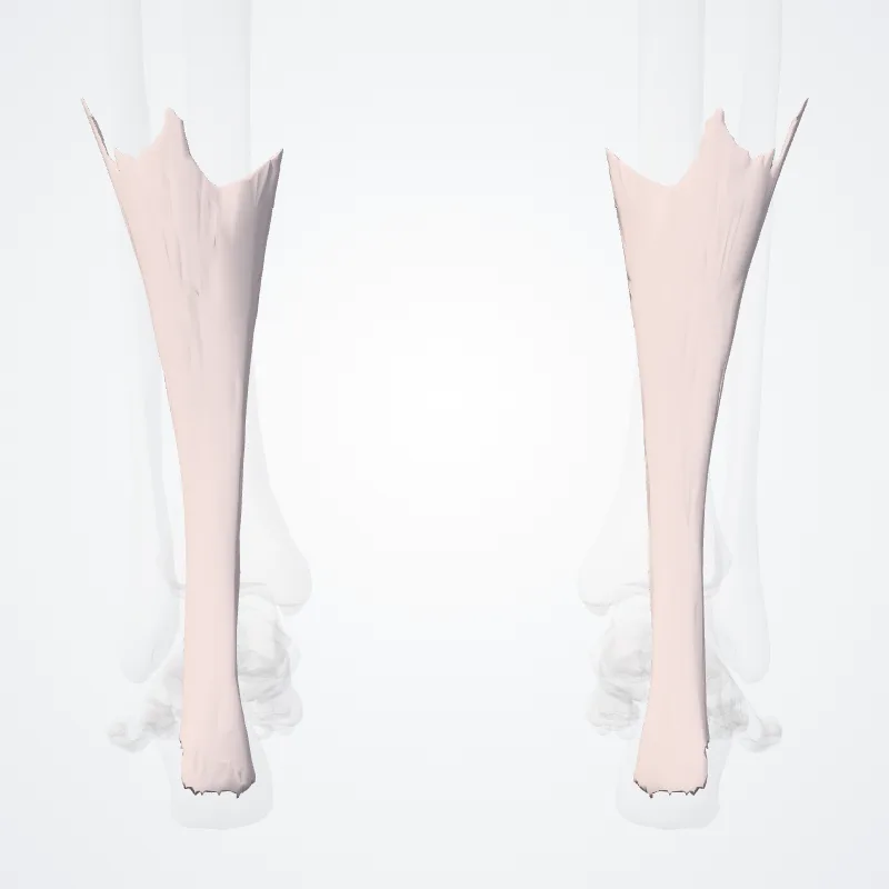
The calcaneal tendon or heel cord, also known as the Achilles tendon, is a tendon at the back of the lower leg, and is the thickest in the human body.
It serves to attach the plantaris, gastrocnemius (calf) and soleus muscles to the calcaneus (heel) bone.
These muscles, acting via the tendon, cause plantar flexion of the foot at the ankle joint, and (except the soleus) flexion at the knee.
Abnormalities of the calcaneal tendon include inflammation (calcaneal tendinitis), degeneration, rupture, and becoming embedded with cholesterol deposits (xanthomas).
The calcaneal tendon was named in 1693 after the Greek hero calcaneal.
Continue reading on Wikipedia →
Source: Wikipedia · Text licence: CC BY-SA 3.0 · This text comes from the source data and has not been reviewed by a person yet.
Names
- Latin
- Tendo calcaneus
- English
- Calcaneal tendon
- French
- Tendon calcanéen
- Spanish
- Tendón calcáneo
- Portuguese
- Tendão calcâneo