Landmark · Skeletal system
Crown of tooth
Corona dentis
- System
- Skeletal system
- Regions
- Head
- Side
- Left and right
- Landmark on
- Lower first molar tooth
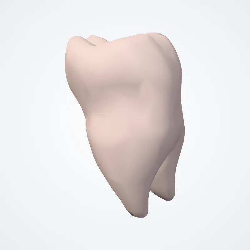
DescriptionUnreviewed
In dentistry, crown refers to the anatomical area of teeth, usually covered by enamel.
The crown is usually visible in the mouth after developing below the gingiva and then erupting into place.
If part of the tooth gets chipped or broken, a dentist can apply an artificial crown. Crowns are used most commonly to entirely cover a damaged tooth or cover an implant.
Bridges are also used to cover a space if one or more teeth is missing.
They are cemented to natural teeth or implants surrounding the space where the tooth once stood. There are various materials that can be used including a type of cement or stainless steel.
The cement crowns look like regular teeth while the stainless steel crowns are silver or gold.
Source: Wikipedia · Text licence: CC BY-SA 3.0 · This text comes from the source data and has not been reviewed by a person yet.
Names
- Latin
- Corona dentis
- English
- Crown of tooth
- French
- Couronne clinique de la dent
- Spanish
- Corona del diente
- Portuguese
- Coroa do dente