Muscular system
Diaphragm
Diaphragma
- System
- Muscular system
- Regions
- Trunk
- Part of
- Thoracic part of muscular system
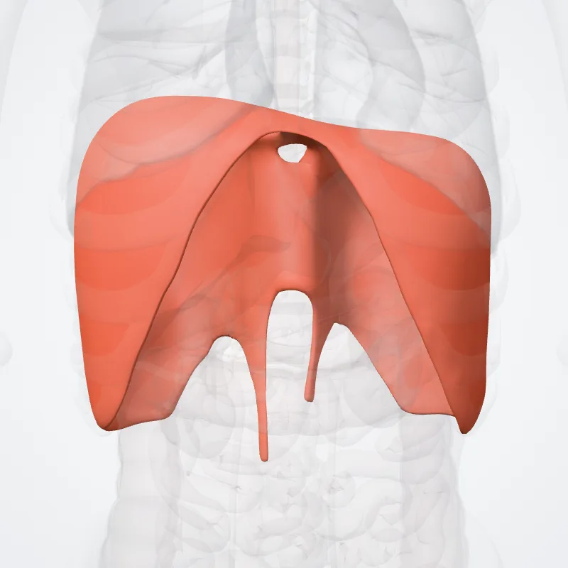
DescriptionUnreviewed
The thoracic diaphragm, or simply the diaphragm (Ancient Greek: διάφραγμα, romanized: diáphragma, lit. 'partition'), is a sheet of internal skeletal muscle in humans and other mammals that extends across the bottom of the thoracic cavity.
The diaphragm is the most important muscle of respiration, and separates the thoracic cavity, containing the heart and lungs, from the abdominal cavity:
as the diaphragm contracts, the volume of the thoracic cavity increases, creating a negative pressure there, which draws air into the lungs.
Its high oxygen consumption is noted by the many mitochondria and capillaries present; more than in any other skeletal muscle.
The term diaphragm in anatomy, created by Gerard of Cremona, can refer to other flat structures such as the urogenital diaphragm or pelvic diaphragm, but "the diaphragm" generally refers to the thoracic diaphragm.
In humans, the diaphragm is slightly asymmetric—its right half is higher up (superior) to the left half, since the large liver rests beneath the right half of the diaphragm.
There is also a theory that the diaphragm is lower on the other side due to the presence of the heart.
Other mammals have diaphragms, and other vertebrates such as amphibians and reptiles have diaphragm-like structures, but important details of the anatomy may vary, such as the position of the lungs in the thoracic cavity.
Continue reading on Wikipedia →
Source: Wikipedia · Text licence: CC BY-SA 3.0 · This text comes from the source data and has not been reviewed by a person yet.
Muscle attachments
Origin
Names
- Latin
- Diaphragma
- English
- Diaphragm
- French
- Diaphragme
- Spanish
- Diafragma
- Portuguese
- Diafragma