Landmark · Skeletal system
Greater horn of hyoid bone
Cornu majus ossis hyoidei
- System
- Skeletal system
- Regions
- Head, Neck, Trunk
- Landmark on
- Hyoid bone
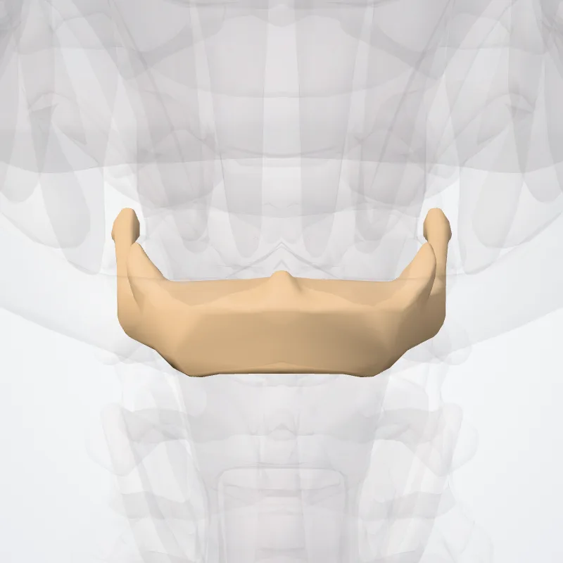
DescriptionUnreviewed
The greater horns project backward from the outer borders of the body; they are flattened from above downward and taper to their end, which is a bony tubercle connecting to the lateral thyrohyoid ligament.
The upper surface of the greater horns are rough and close to its lateral border, and facilitates muscular attachment.
The largest of muscles that attach to the upper surface of the greater horns are the hyoglossus and the middle pharyngeal constrictor, which extend along the whole length of the horns; the digastric muscle and stylohyoid muscle have small insertions in front of these near the junction of the body with the horns.
To the medial border the thyrohyoid membrane is attached, while the anterior half of the lateral border gives insertion to the thyrohyoid muscle.
Source: Wikipedia · Text licence: CC BY-SA 3.0 · This text comes from the source data and has not been reviewed by a person yet.
Names
- Latin
- Cornu majus ossis hyoidei
- English
- Greater horn of hyoid bone
- French
- Grande corne
- Spanish
- Cuerno mayor del hueso hioides
- Portuguese
- Corno maior do osso hióide