Muscular system
Iliotibial tract
Tractus iliotibialis
- System
- Muscular system
- Regions
- Lower limb
- Side
- Left and right
- Part of
- Tensor fasciae latae
DescriptionUnreviewed
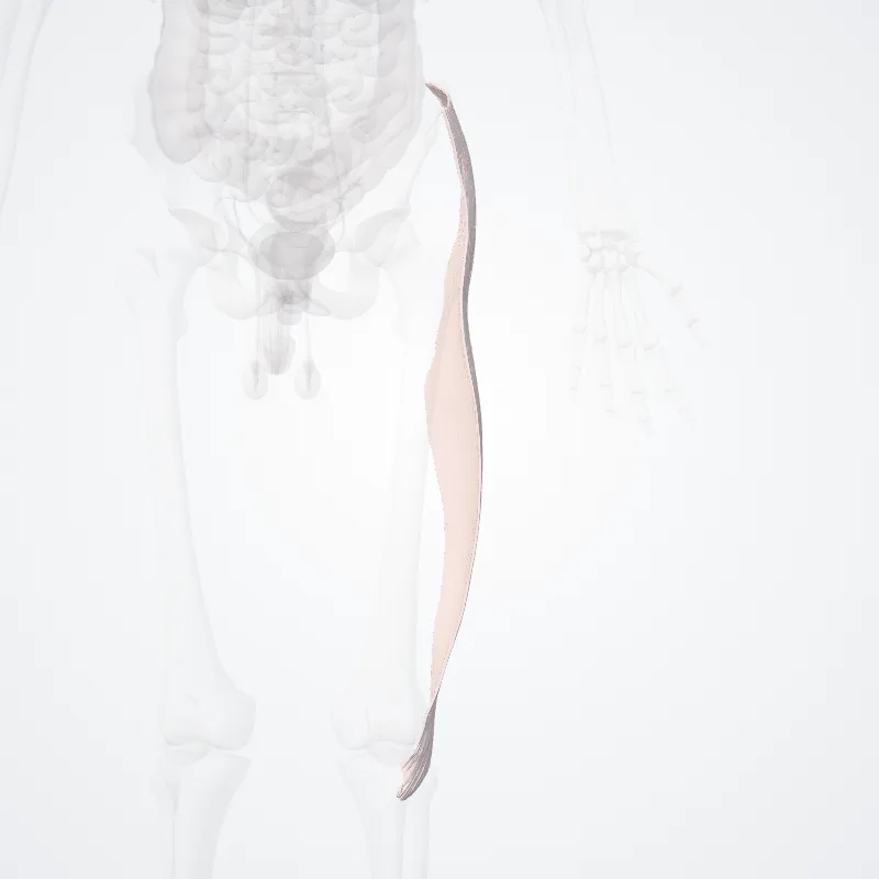
The iliotibial tract or iliotibial band (also known as Maissiat's band or the IT band) is a longitudinal fibrous reinforcement of the fascia lata.
The action of the muscles associated with the ITB (tensor fasciae latae and some fibers of gluteus maximus) flex, extend, abduct, and laterally and medially rotate the hip.
The ITB contributes to lateral knee stabilization.
During knee extension the ITB moves anterior to the lateral condyle of the femur, while ~30 degrees knee flexion, the ITB moves posterior to the lateral condyle.
However, it has been suggested that this is only an illusion due to the changing tension in the anterior and posterior fibers during movement.
It originates at the anterolateral iliac tubercle portion of the external lip of the iliac crest and inserts at the lateral condyle of the tibia at Gerdy's tubercle.
The figure shows only the proximal part of the iliotibial tract.
The part of the iliotibial band which lies beneath the tensor fasciae latae is prolonged upward to join the lateral part of the capsule of the hip-joint.
The tensor fasciae latae effectively tightens the iliotibial band around the area of the knee.
This allows for bracing of the knee especially in lifting the opposite foot.The gluteus maximus muscle and the tensor fasciae latae insert upon the tract.
Continue reading on Wikipedia →
Source: Wikipedia · Text licence: CC BY-SA 3.0 · This text comes from the source data and has not been reviewed by a person yet.
Names
- Latin
- Tractus iliotibialis
- English
- Iliotibial tract
- French
- Tractus iliotibial
- Spanish
- Tracto iliotibial
- Portuguese
- Trato iliotibial