Skeletal system
Major alar cartilage
Cartilago alaris major
- System
- Skeletal system
- Regions
- Head
- Side
- Left and right
- Part of
- Nasal cartilages
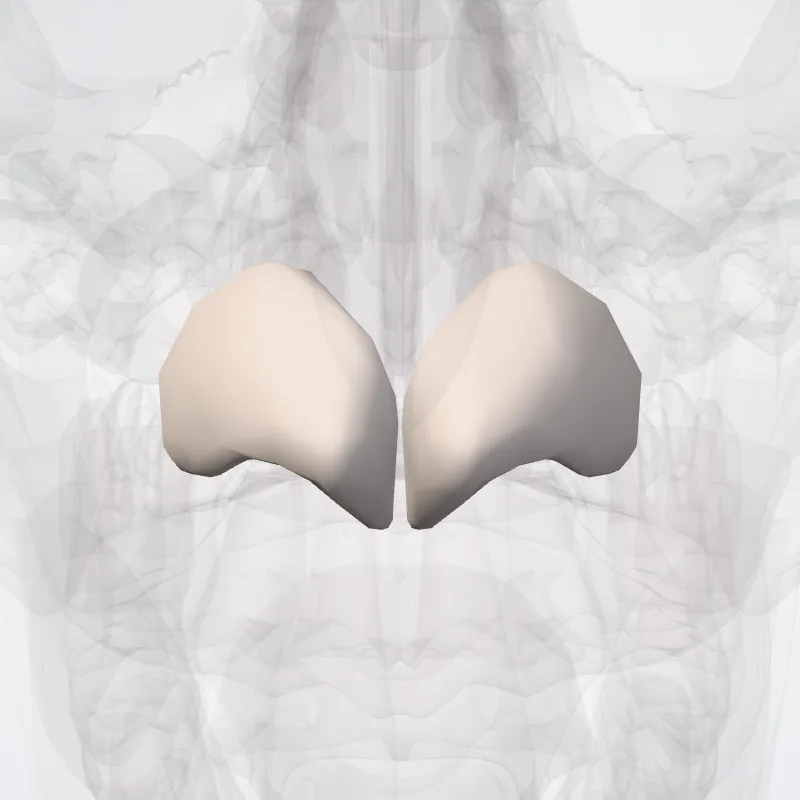
DescriptionUnreviewed
The major alar cartilages are positioned with one structure on each side of the nasal tip.
Superiorly, the major alar cartilages are connected to the lateral nasal cartilage via fibrous tissues.
Composed of hyaline cartilage, these structures are very thin and folded to form the lateral and medial crus.
The medial crus is the inner portion of the major alar cartilages that are situated perpendicularly to the septal nasal cartilage.
The lateral crus is the outer portion of the major alar cartilages that associate with the ala of the nose.
Both crus come together to form an oval tip at each nostril.
Both sides of the major alar cartilages merge together to form a notch at the tip, which is referred to as the apex of the nose.
With the formation of the medial and lateral walls within the nares, the major alar cartilages function to hold open each naris.
This allows maximal airflow to reach the nasal valve, allowing optimal respiration.
Due to weakness corresponding with the lateral crus in certain individuals, a technique called sliding alar cartilage (SAC) has been a procedure practiced to restructure and support the nasal tip.
Source: Z-Anatomy · Text licence: CC BY-SA 4.0 · This text comes from the source data and has not been reviewed by a person yet.
Names
- Latin
- Cartilago alaris major
- English
- Major alar cartilage
- French
- Grand cartilage alaire
- Spanish
- Cartílago alar mayor
- Portuguese
- Cartilagem alar maior