Cardiovascular system
Middle meningeal artery
Arteria meningea media
- System
- Cardiovascular system
- Regions
- Head
- Side
- Left and right
- Part of
- Maxillary artery
DescriptionUnreviewed
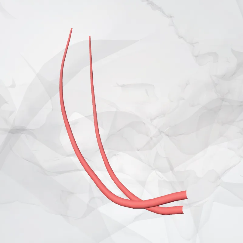
The middle meningeal artery (Latin: arteria meningea media) is typically the third branch of the first portion of the maxillary artery.
After branching off the maxillary artery in the infratemporal fossa, it runs through the foramen spinosum to supply the dura mater (the outer meningeal layer) and the calvaria.
The middle meningeal artery is the largest of the three (paired) arteries that supply the meninges, the others being the anterior meningeal artery and the posterior meningeal artery. The anterior branch of the middle meningeal artery runs beneath the pterion.
It is vulnerable to injury at this point, where the skull is thin.
Rupture of the artery may give rise to an epidural hematoma.
In the dry cranium, the middle meningeal, which runs within the dura mater surrounding the brain, makes a deep groove in the calvarium. The middle meningeal artery is intimately associated with the auriculotemporal nerve, which wraps around the artery making the two easily identifiable in the dissection of human cadavers and also easily damaged in surgery.
Continue reading on Wikipedia →
Source: Wikipedia · Text licence: CC BY-SA 3.0 · This text comes from the source data and has not been reviewed by a person yet.
Parts
Names
- Latin
- Arteria meningea media
- English
- Middle meningeal artery
- French
- Artère méningée moyenne
- Spanish
- Arteria meníngea media
- Portuguese
- Artéria meníngea média
In the same group
- Anterior deep temporal artery
- Artery of pterygoid canal
- Buccal artery
- Descending palatine artery
- Inferior alveolar artery
- Infra-orbital artery
- Posterior deep temporal artery
- Posterior lateral nasal branches of sphenopalatine artery.
- Posterior septal branches of sphenopalatine artery
- Posterior superior alveolar artery