Group · Muscular system
Pharyngeal muscles
Musculi pharyngis
- System
- Muscular system
- Regions
- Neck, Trunk
- Part of
- Muscles of neck
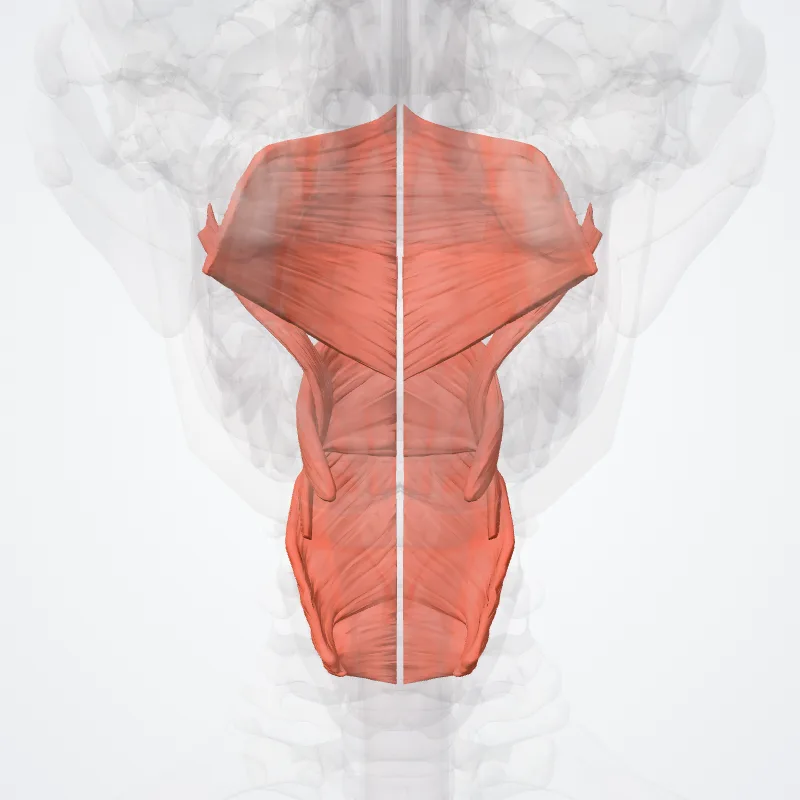
DescriptionUnreviewed
The pharyngeal muscles are a group of muscles that form the pharynx, which is posterior to the oral cavity, determining the shape of its lumen, and affecting its sound properties as the primary resonating cavity.
The pharyngeal muscles (involuntary skeletal) push food into the esophagus.
There are two muscular layers of the pharynx: the outer circular layer and the inner longitudinal layer.
The outer circular layer includes:
- Inferior constrictor muscle
- Middle constrictor muscle
- Superior constrictor muscle
During swallowing, these muscles constrict to propel a bolus downwards (an involuntary process).
The inner longitudinal layer includes:
- Stylopharyngeus muscle
- Salpingopharyngeus muscle
- Palatopharyngeus muscleDuring swallowing, these muscles act to shorten and widen the pharynx.
They are innervated by the pharyngeal branch of the vagus nerve (CN X) with the exception of the stylopharyngeus muscle which is innervated by the glossopharyngeal nerve (CN IX).
Source: Wikipedia · Text licence: CC BY-SA 3.0 · This text comes from the source data and has not been reviewed by a person yet.
Parts
Names
- Latin
- Musculi pharyngis
- English
- Pharyngeal muscles
- French
- Muscles pharyngiens
- Spanish
- Músculos faríngeos
- Portuguese
- Músculos faríngeos