Joints & ligaments
Posterior cruciate ligament
Ligamentum decussatum posterius
- System
- Joints & ligaments
- Regions
- Lower limb
- Side
- Left and right
- Part of
- Knee joint
DescriptionUnreviewed
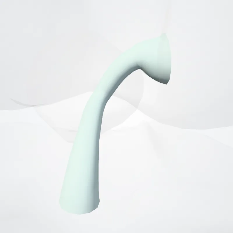
The posterior cruciate ligament (PCL) is a ligament in each knee of humans and various other animals.
It works as a counterpart to the anterior cruciate ligament (ACL).
It connects the posterior intercondylar area of the tibia to the medial condyle of the femur.
This configuration allows the PCL to resist forces pushing the tibia posteriorly relative to the femur. The PCL and ACL are intracapsular ligaments because they lie deep within the knee joint.
They are both isolated from the fluid-filled synovial cavity, with the synovial membrane wrapped around them.
The PCL gets its name by attaching to the posterior portion of the tibia.The PCL, ACL, MCL, and LCL are the four main ligaments of the knee in primates.
Continue reading on Wikipedia →
Source: Wikipedia · Text licence: CC BY-SA 3.0 · This text comes from the source data and has not been reviewed by a person yet.
Names
- Latin
- Ligamentum decussatum posterius
- English
- Posterior cruciate ligament
- French
- Ligament croisé postérieur
- Spanish
- Ligamento cruzado posterior
- Portuguese
- Ligamento cruzado posterior
In the same group
- Anterior cruciate ligament
- Anterior meniscotibial ligament (Lateral meniscus)
- Anterior meniscotibial ligament (Medial meniscus)
- Arcuate popliteal ligament
- Articular capsule of knee joint
- Deep part of tibial collateral ligament
- Fibular collateral ligament
- Infrapatellar fat pad
- Lateral meniscus
- Medial meniscus
- Meniscopatellar ligament
- Oblique popliteal ligament
- Popliteofibular ligament
- Posterior meniscotibial ligament (Lateral meniscus)
- Posterior meniscotibial ligament (Medial meniscus)
- Superficial part of tibial collateral ligament
- Transverse ligament of knee