Landmark · Skeletal system
Posterior surface of scapula
Facies posterior scapulae
- System
- Skeletal system
- Regions
- Trunk, Upper limb
- Side
- Left and right
- Landmark on
- Scapula
DescriptionUnreviewed
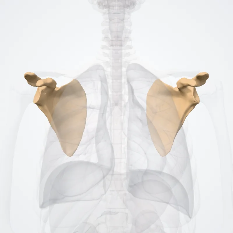
The back of the scapula (also called the dorsal or posterior surface) is arched from above downward, and is subdivided into two unequal parts by the spine of the scapula.
The portion above the spine is called the supraspinous fossa, and that below it the infraspinous fossa.
The two fossae are connected by the spinoglenoid notch, situated lateral to the root of the spine.
The supraspinous fossa, above the spine of scapula, is concave, smooth, and broader at its vertebral than at its humeral end; its medial two-thirds give origin to the Supraspinatus.
At its lateral surface resides the spinoglenoid fossa which is situated by the medial margin of the glenoid.
The spinoglenoid fossa houses the suprascpular canal which forms a connecting passage between the suprascapular notch and the spinoglenoid notch conveying the suprascapular nerve and vessels.
The infraspinous fossa is much larger than the preceding; toward its vertebral margin a shallow concavity is seen at its upper part; its center presents a prominent convexity, while near the axillary border is a deep groove which runs from the upper toward the lower part.
The medial two-thirds of the fossa give origin to the Infraspinatus; the lateral third is covered by this muscle.
There is a ridge on the outer part of the back of the scapula.
This runs from the lower part of the glenoid cavity, downward and backward to the vertebral border, about 2.5 cm above the inferior angle.
Attached to the ridge is a fibrous septum, which separates the infraspinatus muscle from the Teres major and Teres minor muscles.
The upper two-thirds of the surface between the ridge and the axillary border is narrow, and is crossed near its center by a groove for the scapular circumflex vessels; the Teres minor attaches here.
The broad and narrow portions above alluded to are separated by an oblique line, which runs from the axillary border, downward and backward, to meet the elevated ridge: to it is attached a fibrous septum which separates the Teres muscles from each other.
Its lower third presents a broader, somewhat triangular surface, the inferior angle of the scapula, which gives origin to the Teres major, and over which the Latissimus dorsi glides; frequently the latter muscle takes origin by a few fibers from this part.
Source: Z-Anatomy · Text licence: CC BY-SA 4.0 · This text comes from the source data and has not been reviewed by a person yet.
Names
- Latin
- Facies posterior scapulae
- English
- Posterior surface of scapula
- French
- Face postérieure de la scapula
- Spanish
- Cara posterior de la escápula
- Portuguese
- Superfície posterior da escápula