Landmark · Skeletal system
Ramus of ischium
Ramus ossis ischii
- System
- Skeletal system
- Regions
- Trunk, Pelvis, Lower limb
- Side
- Left and right
- Landmark on
- Hip bone
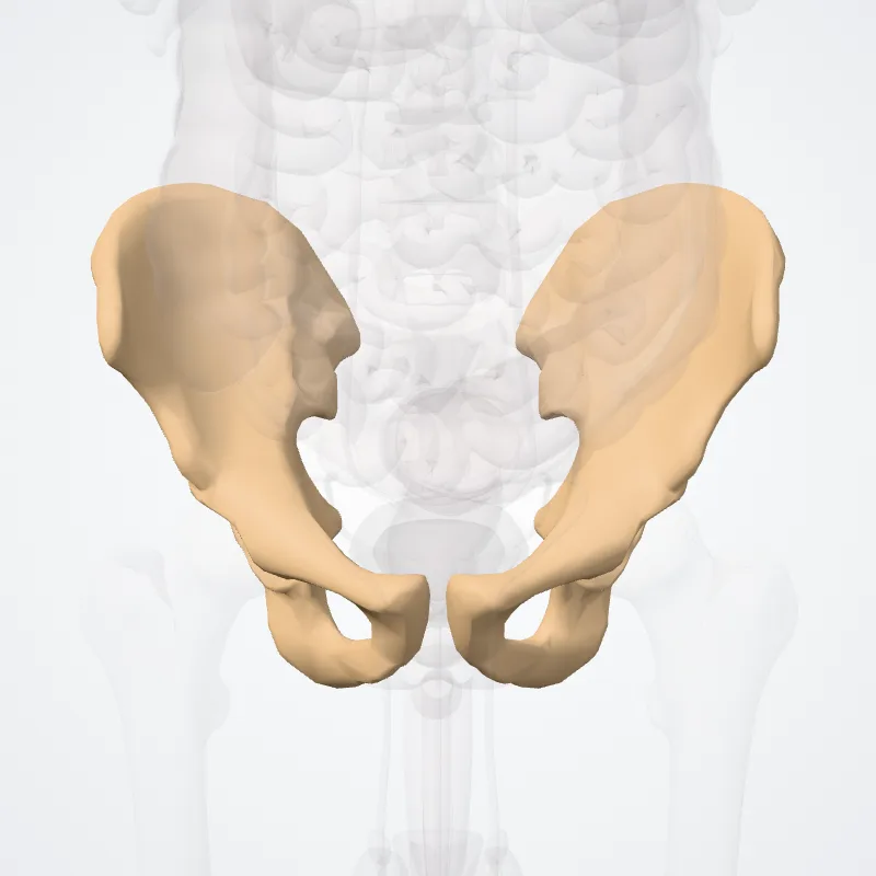
DescriptionUnreviewed
Superior ramus
The superior ramus of the ischium (descending ramus) projects downward and backward from the body and presents for examination three surfaces: external, internal, and posterior.
The external surface is quadrilateral in shape.
It is bounded above by a groove that lodges the tendon of the external obturator; below, it is continuous with the inferior ramus; in front it is limited by the posterior margin of the obturator foramen; behind, a prominent margin separates it from the posterior surface.
In front of this margin the surface gives origin to the quadratus femoris, and anterior to this to some of the fibers of origin of the external obturator; the lower part of the surface gives origin to part of the adductor magnus.
The internal surface forms part of the bony wall of the lesser pelvis. In front it is limited by the posterior margin of the obturator foramen. Below, it is bounded by a sharp ridge that provides attachment to a falciform prolongation of the sacrotuberous ligament, and, more anteriorly, gives origin to the transverse perineal and ischiocavernosus muscles.
Posteriorly the ramus forms a large swelling, the tuberosity of the ischium, where the hamstrings originate.
Inferior ramus
The inferior ramus of the ischium (ascending ramus) is the thin, flattened part of the ischium, which ascends from the superior ramus, and joins the inferior ramus of the pubis—the junction being indicated in the adult by a raised line.
The outer surface is uneven for the origin of the obturator externus and some of the fibers of the adductor magnus; its inner surface forms part of the anterior wall of the pelvis.
Its medial border is thick, rough, slightly everted, forms part of the outlet of the pelvis, and presents two ridges and an intervening space.
The ridges are continuous with similar ones on the inferior ramus of the pubis: to the outer is attached the deep layer of the superficial perineal fascia (fascia of Colles), and to the inner the inferior fascia of the urogenital diaphragm.
Tracing these two ridges downward, they join with each other just behind the point of origin of the transverse perineal muscles.
Here the two layers of fascia are continuous behind the posterior border of the muscle.
To the intervening space, just in front of the point of junction of the ridges, the transverse perineal attaches, and in front of this is a portion of the ischiocavernosus, and the crus penis in the male, or the crus clitoridis in the female.
Its lateral border is thin and sharp, and forms part of the medial margin of the obturator foramen.
Source: Z-Anatomy · Text licence: CC BY-SA 4.0 · This text comes from the source data and has not been reviewed by a person yet.
Names
- Latin
- Ramus ossis ischii
- English
- Ramus of ischium
- French
- Branche de l'ischium
- Spanish
- Ramo del hueso isquion
- Portuguese
- Ramo de ísquio