Muscular system
Rectus abdominis muscle
Musculus rectus abdominis
- System
- Muscular system
- Regions
- Trunk
- Side
- Left and right
- Part of
- Muscles of abdomen
DescriptionUnreviewed
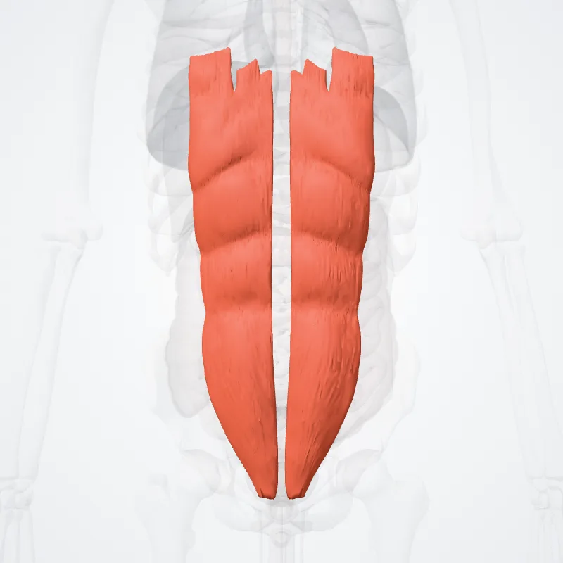
The rectus abdominis muscle, also known as the "abdominal muscle", is a paired muscle running vertically on each side of the anterior wall of the human abdomen, as well as that of some other mammals.
There are two parallel muscles, separated by a midline band of connective tissue called the linea alba.
It extends from the pubic symphysis, pubic crest and pubic tubercle inferiorly, to the xiphoid process and costal cartilages of ribs V to VII superiorly.
The proximal attachments are the pubic crest and the pubic symphysis.
It attaches distally at the costal cartilages of ribs 5-7 and the xiphoid process of the sternum.The rectus abdominis muscle is contained in the rectus sheath, which consists of the aponeuroses of the lateral abdominal muscles.
The outer, most lateral line, defining the rectus is the linea semilunaris.
Bands of connective tissue traverse the rectus abdominis, separating it into distinct muscle bellies.
In the abdomens of people with low body fat, these muscle bellies can be viewed externally.
They can appear in sets of as few as two to as many as twelve.
Although, six is the most common.
Continue reading on Wikipedia →
Source: Wikipedia · Text licence: CC BY-SA 3.0 · This text comes from the source data and has not been reviewed by a person yet.
Muscle attachments
Origin
Insertion
Names
- Latin
- Musculus rectus abdominis
- English
- Rectus abdominis muscle
- French
- Mucle droit de l'abdomen
- Spanish
- Músculo recto del abdomen
- Portuguese
- Músculo reto abdominal