Nervous system & sense organs
Suspensory ligament of eyeball
Ligamentum suspensorium bulbi
- System
- Nervous system & sense organs
- Regions
- Head
- Side
- Left and right
- Part of
- Accessory visual structures
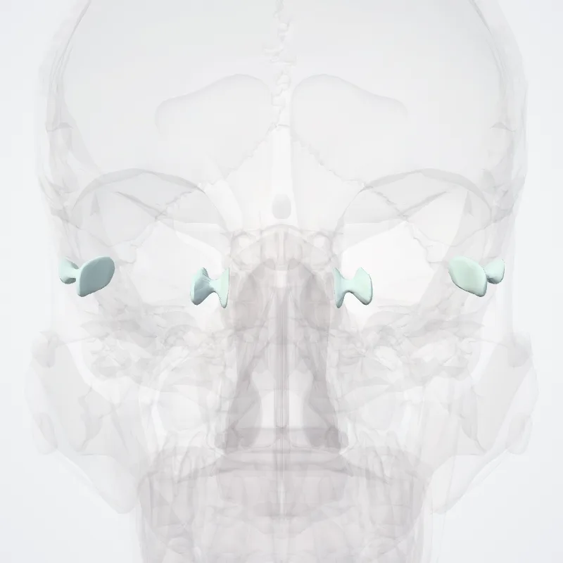
DescriptionUnreviewed
The suspensory ligament of eyeball (or Lockwood's ligament) forms a hammock stretching below the eyeball between the medial and lateral check ligaments and enclosing the inferior rectus and inferior oblique muscles of the eye.
It is a thickening of Tenon's capsule, the dense connective tissue capsule surrounding the globe and separating it from orbital fat.
This ligament is responsible for maintaining and supporting the position of the eyeball in its normal upward and forward position within the orbit, and prevents downward displacement of the eyeball.
It can be considered a part of the bulbar sheath.It is named for Charles Barrett Lockwood.
Source: Wikipedia · Text licence: CC BY-SA 3.0 · This text comes from the source data and has not been reviewed by a person yet.
Names
- Latin
- Ligamentum suspensorium bulbi
- English
- Suspensory ligament of eyeball
- French
- Ligament suspenseur du bulbe
- Spanish
- Ligamento suspensorio del globo ocular
- Portuguese
- Ligamento suspensor do globo ocular