Group · Joints & ligaments
Synovial joints of thorax
Articulationes thoracis
- System
- Joints & ligaments
- Regions
- Trunk, Thorax
- Part of
- Thoracic joints
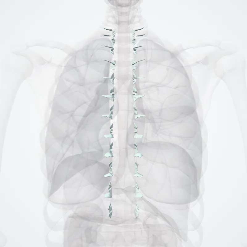
DescriptionUnreviewed
A synovial joint, also known as diarthrosis, joins bones or cartilage with a fibrous joint capsule that is continuous with the periosteum of the joined bones, constitutes the outer boundary of a synovial cavity, and surrounds the bones' articulating surfaces.
This joint unites long bones and permits free bone movement and greater mobility.
The synovial cavity/joint is filled with synovial fluid.
The joint capsule is made up of an outer layer, the articular capsule, which keeps the bones together structurally, and an inner layer, the synovial membrane, which seals in the synovial fluid.
They are the most common and most movable type of joint in the body of a mammal.
As with most other joints, synovial joints achieve movement at the point of contact of the articulating bones.
Continue reading on Wikipedia →
Source: Wikipedia · Text licence: CC BY-SA 3.0 · This text comes from the source data and has not been reviewed by a person yet.
Parts
Names
- Latin
- Articulationes thoracis
- English
- Synovial joints of thorax
- French
- Articulations synoviales du thorax
- Spanish
- Articulaciones sinoviales del tórax
- Portuguese
- Articulações sinoviais do tórax