Skeletal system
Talus
Os tali
- System
- Skeletal system
- Regions
- Lower limb, Foot
- Side
- Left and right
- Part of
- Tarsal bones
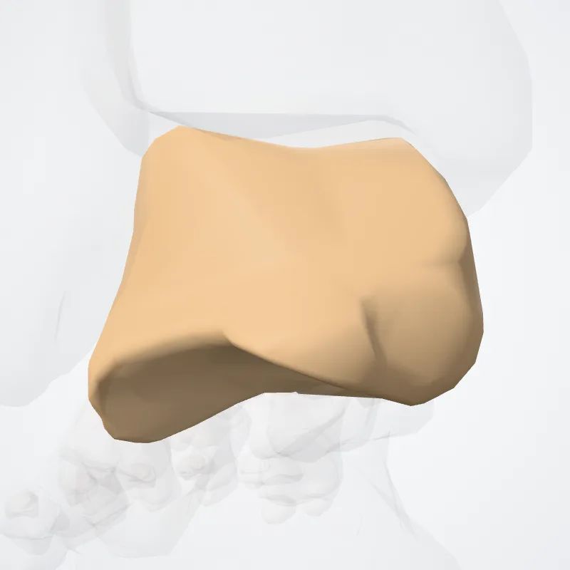
DescriptionUnreviewed
The talus (Latin for ankle), talus bone, astragalus, or ankle bone is one of the group of foot bones known as the tarsus.
The tarsus forms the lower part of the ankle joint.
It transmits the entire weight of the body from the lower legs to the foot.
The talus has joints with the two bones of the lower leg, the tibia and thinner fibula.
These leg bones have two prominences (the lateral and medial malleoli) that articulate with the talus.
At the foot end, within the tarsus, the talus articulates with the calcaneus (heel bone) below, and with the curved navicular bone in front; together, these foot articulations form the ball-and-socket-shaped talocalcaneonavicular joint.
The talus is the second largest of the tarsal bones; it is also one of the bones in the human body with the highest percentage of its surface area covered by articular cartilage.
It is also unusual in that it has a retrograde blood supply, i.e. arterial blood enters the bone at the distal end.
In humans, no muscles attach to the talus, unlike most bones, and its position therefore depends on the position of the neighbouring bones.
Continue reading on Wikipedia →
Source: Wikipedia · Text licence: CC BY-SA 3.0 · This text comes from the source data and has not been reviewed by a person yet.
Landmarks
- Body of talus
- Facet for calcaneonavicular part of bifurcate ligament
- Facet for plantar calcaneonavicular ligament
- Groove for tendon of flexor hallucis longus of talus
- Head of talus
- Lateral malleolar facet
- Lateral process of talus
- Lateral tubercle
- Medial malleolar facet
- Medial tubercle
- Middle facet for calcaneus
- Navicular articular surface
- Neck of talus
- Posterior calcaneal articular facet
- Posterior process of talus
- Superior facet of talus
- Talar sulcus
- Trochlea of talus
Names
- Latin
- Os tali
- English
- Talus
- French
- Talus
- Spanish
- Hueso Talo
- Portuguese
- Talus