Group · Joints & ligaments
Tibiofibular syndesmosis
Syndesmosis tibiofibularis
- System
- Joints & ligaments
- Regions
- Lower limb, Foot
- Part of
- Fibrous joints of free lower limb
DescriptionUnreviewed
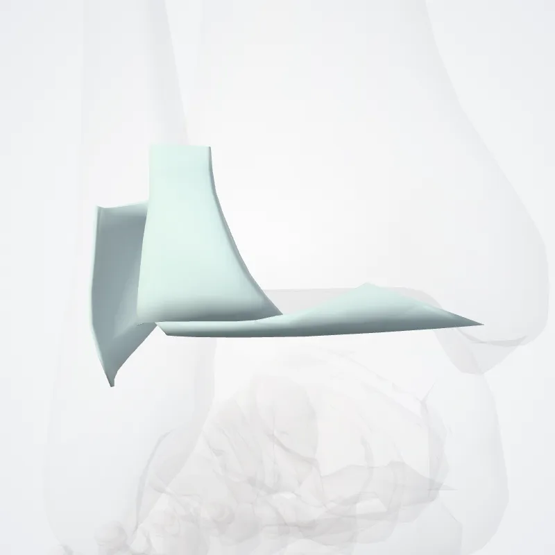
The distal tibiofibular joint is a syndesmosis; it is formed by the rough, convex surface of the medial side of the distal end of the fibula, and a rough concave surface on the lateral side of the tibia.
Below, to the extent of about 4 mm, these surfaces are smooth and covered with cartilage, which is continuous with that of the ankle joint.
The ligaments are:
Anterior ligament of the lateral malleolus Posterior ligament of the lateral malleolus Interosseous membrane of leg
The inferior transverse ligament of the tibiofibular syndesmosis is included in older versions of Gray's Anatomy, but not in Terminologia Anatomica.
However, it still appears in some anatomy textbooks.
It should not be confused with the superior tibiofibular joint, which is the only synovial tibiofibular joint, and is sometimes simply called the "tibiofibular articulation".
Source: Wikipedia · Text licence: CC BY-SA 3.0 · This text comes from the source data and has not been reviewed by a person yet.
Parts
Names
- Latin
- Syndesmosis tibiofibularis
- English
- Tibiofibular syndesmosis
- French
- Syndesmose tibiofibulaire
- Spanish
- Sindesmosis tibiofibular
- Portuguese
- Sindesmose tibiofibular