Landmark · Skeletal system
Tuberosity of distal phalanx of foot
Tuberositas phalangis distalis pedis
- System
- Skeletal system
- Regions
- Lower limb, Foot
- Side
- Left and right
- Landmark on
- Distal phalanx of third finger of foot
DescriptionUnreviewed
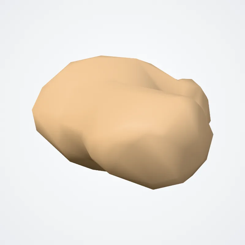
The phalanx ends in an expanded distal extremity for the support of the nail and end of the toe; it is a crescent-shaped rough cap of bone epiphysis — the apical tuft (or ungual tuberosity/process) which covers a larger portion of the phalanx on the volar side than on the dorsal side.
Two lateral ungual spines project proximally from the apical tuft.
Near the base of the shaft are two lateral tubercles.
Between these a V-shaped ridge extending proximally serves for the insertion of the flexor pollicis longus.
Another ridge at the base serves for the insertion of the extensor aponeurosis.
The flexor insertion is sided by two fossae — the ungual fossa distally and the proximopalmar fossa proximally.
Source: Wikipedia · Text licence: CC BY-SA 3.0 · This text comes from the source data and has not been reviewed by a person yet.
Names
- Latin
- Tuberositas phalangis distalis pedis
- English
- Tuberosity of distal phalanx of foot
- French
- Tubérosité de la phalange distale du pied
- Spanish
- Tuberosidad de la falange distal del pie
- Portuguese
- Tuberosidade da falange distal do pé