Nervensystem und Sinnesorgane
Ventriculus lateralis
Lateral ventricle
- System
- Nervensystem und Sinnesorgane
- Regionen
- Kopf
- Seite
- Links und rechts
- Teil von
- Telencephalon
BeschreibungUngeprüft
Beschreibungen gibt es nur auf Englisch.
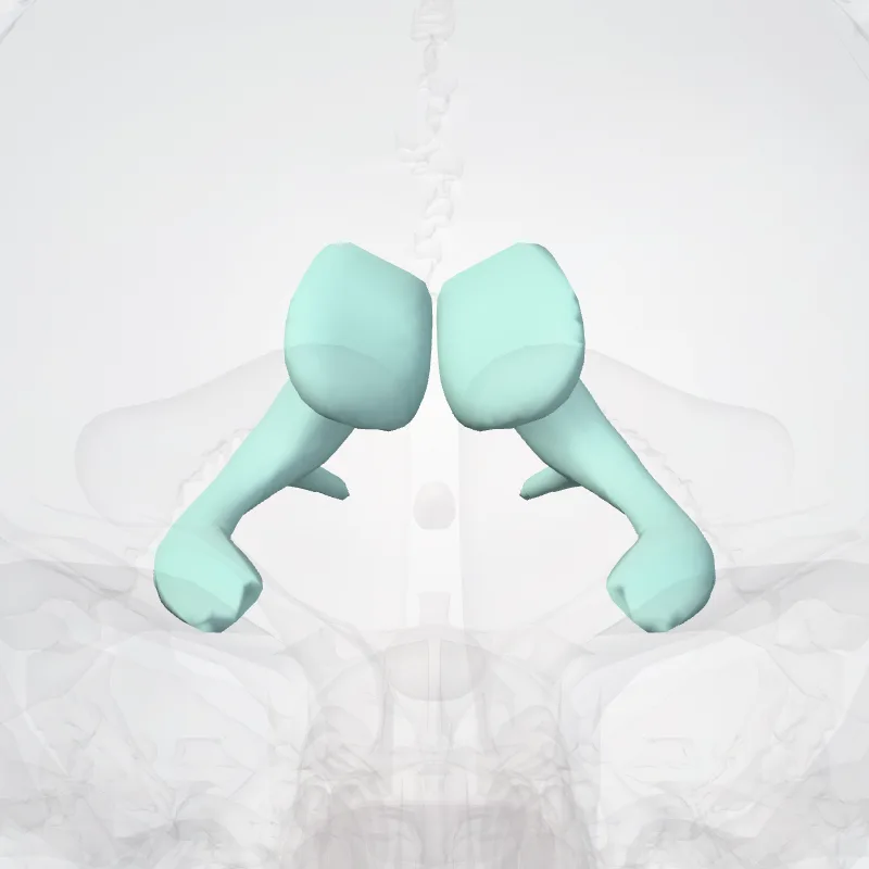
The lateral ventricles are the two largest ventricles of the brain and contain cerebrospinal fluid (CSF).
Each cerebral hemisphere contains a lateral ventricle, known as the left or right ventricle, respectively.
Each lateral ventricle resembles a C-shaped cavity that begins at an inferior horn in the temporal lobe, travels through a body in the parietal lobe and frontal lobe, and ultimately terminates at the interventricular foramina where each lateral ventricle connects to the single, central third ventricle.
Along the path, a posterior horn extends backward into the occipital lobe, and an anterior horn extends farther into the frontal lobe.
Quelle: Wikipedia · Textlizenz: CC BY-SA 3.0 · Dieser Text stammt aus den Quelldaten und wurde noch nicht von einer Person geprüft.
Namen
- Latein
- Ventriculus lateralis
- Englisch
- Lateral ventricle
- Französisch
- Ventricule latéral
- Spanisch
- Ventrículo lateral
- Portugiesisch
- Ventrículo lateral