Nervous system & sense organs
Lateral ventricle
Ventriculus lateralis
- System
- Nervous system & sense organs
- Regions
- Head
- Side
- Left and right
- Part of
- Telencephalon
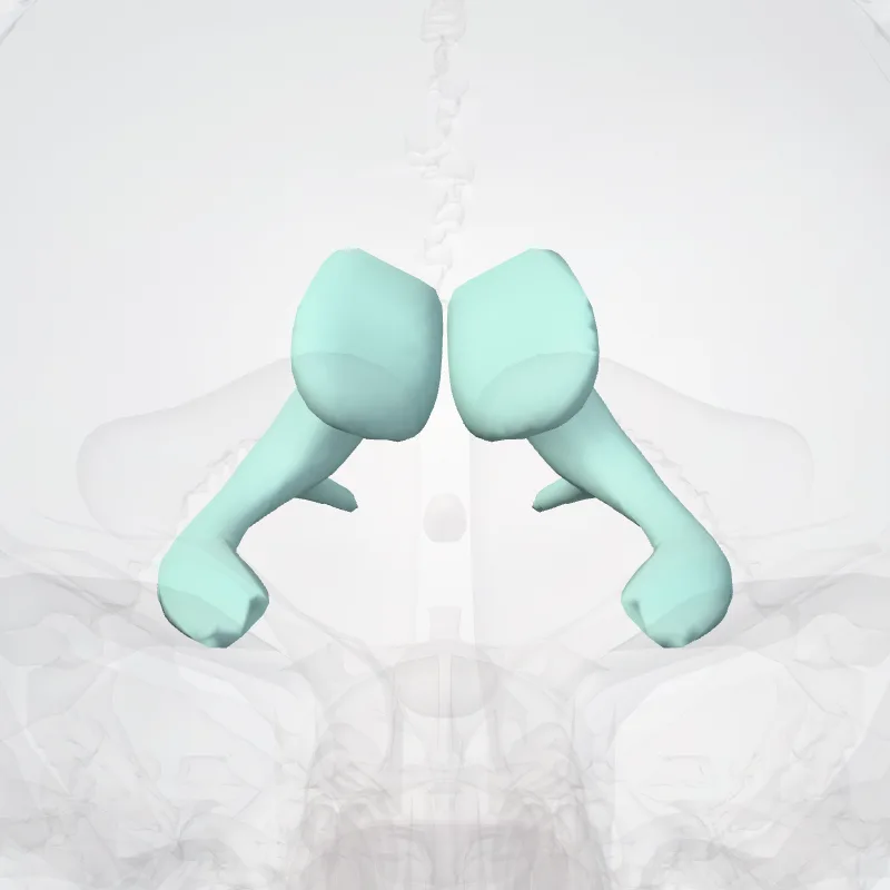
DescriptionUnreviewed
The lateral ventricles are the two largest ventricles of the brain and contain cerebrospinal fluid (CSF).
Each cerebral hemisphere contains a lateral ventricle, known as the left or right ventricle, respectively.
Each lateral ventricle resembles a C-shaped cavity that begins at an inferior horn in the temporal lobe, travels through a body in the parietal lobe and frontal lobe, and ultimately terminates at the interventricular foramina where each lateral ventricle connects to the single, central third ventricle.
Along the path, a posterior horn extends backward into the occipital lobe, and an anterior horn extends farther into the frontal lobe.
Continue reading on Wikipedia →
Source: Wikipedia · Text licence: CC BY-SA 3.0 · This text comes from the source data and has not been reviewed by a person yet.
Names
- Latin
- Ventriculus lateralis
- English
- Lateral ventricle
- French
- Ventricule latéral
- Spanish
- Ventrículo lateral
- Portuguese
- Ventrículo lateral