Landmark · Skeletal system
Medial malleolus
Malleolus medialis
- System
- Skeletal system
- Regions
- Lower limb
- Side
- Left and right
- Landmark on
- Tibia
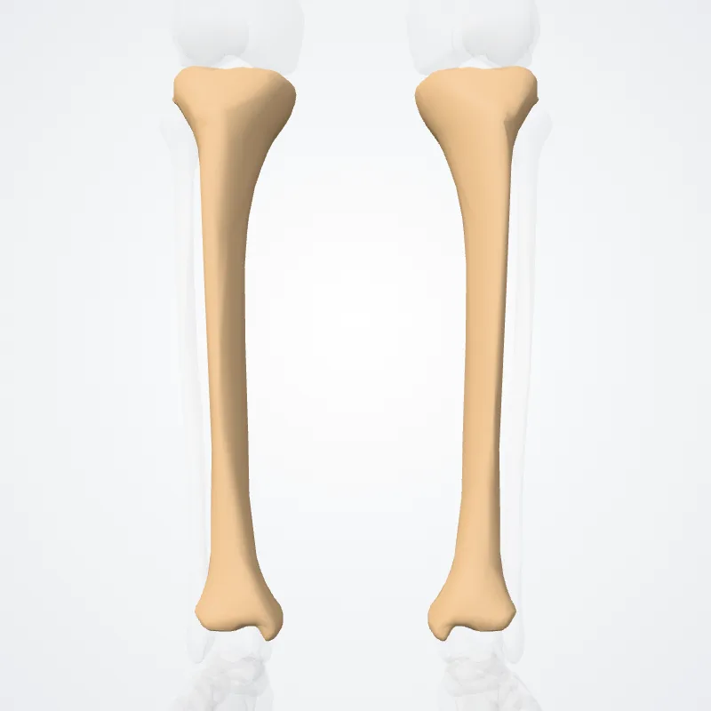
DescriptionUnreviewed
The medial malleolus is found at the foot end of the tibia.
The medial surface of the lower extremity of tibia is prolonged downward to form a strong pyramidal process, flattened from without inward - the medial malleolus.
The medial surface of this process is convex and subcutaneous.
The lateral or articular surface is smooth and slightly concave, and articulates with the talus.
The anterior border is rough, for the attachment of the anterior fibers of the deltoid ligament of the ankle-joint.
The posterior border presents a broad groove, the malleolar sulcus, directed obliquely downward and medially, and occasionally double; this sulcus lodges the tendons of the Tibialis posterior and Flexor digitorum longus.
The summit of the medial malleolus is marked by a rough depression behind, for the attachment of the deltoid ligament.
The major structure that passes anterior to the medial mallelous is the saphenous vein.
Structures that pass behind medial malleolus deep to the flexor retinaculum:
- Tibialis posterior tendon
- Flexor digitorum longus
- Posterior tibial artery
- Posterior tibial vein
- Tibial nerve
- Flexor hallucis longus
Source: Z-Anatomy · Text licence: CC BY-SA 4.0 · This text comes from the source data and has not been reviewed by a person yet.
Names
- Latin
- Malleolus medialis
- English
- Medial malleolus
- French
- Malléole médiale
- Spanish
- Maléolo medial
- Portuguese
- Maléolo medial