Cardiovascular system
Patellar anastomosis
Rete patellare
- System
- Cardiovascular system
- Regions
- Lower limb
- Side
- Left and right
- Part of
- Popliteal artery
DescriptionUnreviewed
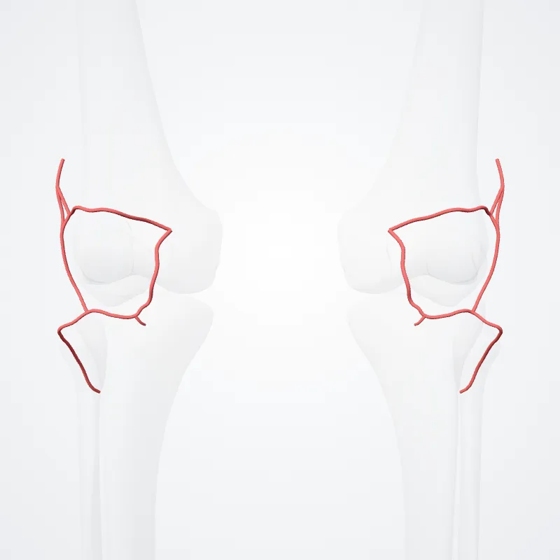
The patellar network (circulatory anastomosis around the knee-joint, patellar anastomosis, genicular anastomosis, articular vascular network of knee or rete articulare genus) is an intricate network of blood vessels around and above the patella, and on the contiguous ends of the femur and tibia, forming a superficial and a deep plexus.
The superficial plexus is situated between the fascia and skin around about the patella, and forms three well-defined arches: one, above the upper border of the patella, in the loose connective tissue over the Quadriceps femoris; the other two, below the level of the patella, are situated in the fat behind the ligamentum patellæ.
The deep plexus, which forms a close net-work of vessels, lies on the lower end of the femur and upper end of the tibia around their articular surfaces, and sends numerous offsets into the interior of the joint.
The arteries which form this plexus are the inferior medial and superior medial genicular arteries, the inferior lateral and superior lateral genicular arteries, the descending genicular artery, the descending branch of lateral femoral circumflex artery, and the anterior tibial recurrent artery.
Continue reading on Wikipedia →
Source: Wikipedia · Text licence: CC BY-SA 3.0 · This text comes from the source data and has not been reviewed by a person yet.
Names
- Latin
- Rete patellare
- English
- Patellar anastomosis
- French
- Réseau patellaire
- Spanish
- Anastomosis patelar
- Portuguese
- Anastomose patelar