Landmark · Skeletal system
Posterior intercondylar area
Area intercondylaris posterior
- System
- Skeletal system
- Regions
- Lower limb
- Side
- Left and right
- Landmark on
- Tibia
DescriptionUnreviewed
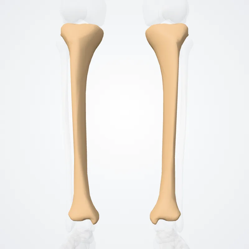
Posteriorly, the condyles are separated from each other by a shallow depression, the posterior intercondyloid fossa, which gives attachment to part of the posterior cruciate ligament of the knee-joint.
The medial condyle presents posteriorly a deep transverse groove, for the insertion of the tendon of the semimembranosus.
Source: Z-Anatomy · Text licence: CC BY-SA 4.0 · This text comes from the source data and has not been reviewed by a person yet.
Names
- Latin
- Area intercondylaris posterior
- English
- Posterior intercondylar area
- French
- Aire intecondylaire postérieure
- Spanish
- Área intercondilar posterior
- Portuguese
- Área intercondilar posterior