Group · Lymphoid organs
Secondary lymphoid organs
Organa lymphoidea secundaria
- System
- Lymphoid organs
- Regions
- Trunk, Head, Lower limb, Neck, Upper limb
DescriptionUnreviewed
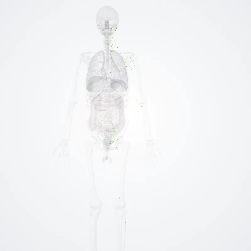
The secondary (or peripheral) lymphoid organs (SLO), which include lymph nodes and the spleen, maintain mature naive lymphocytes and initiate an adaptive immune response.
The secondary lymphoid organs are the sites of lymphocyte activation by antigens.
Activation leads to clonal expansion and affinity maturation.
Mature lymphocytes recirculate between the blood and the secondary lymphoid organs until they encounter their specific antigen.
- SPLEEN:
The main functions of the spleen are:
- to produce immune cells to fight antigens
- to remove particulate matter and aged blood cells, mainly red blood cells
- to produce blood cells during fetal life.
The spleen synthesizes antibodies in its white pulp and removes antibody-coated bacteria and antibody-coated blood cells by way of blood and lymph node circulation.
A study published in 2009 using mice found that the spleen contains, in its reserve, half of the body's monocytes within the red pulp.
These monocytes, upon moving to injured tissue (such as the heart), turn into dendritic cells and macrophages while promoting tissue healing.
The spleen is a center of activity of the mononuclear phagocyte system and can be considered analogous to a large lymph node, as its absence causes a predisposition to certain infections.
Like the thymus, the spleen has only efferent lymphatic vessels. Both the short gastric arteries and the splenic artery supply it with blood.
The germinal centers are supplied by arterioles called penicilliary radicles.
Until the fifth month of prenatal development, the spleen creates red blood cells; after birth, the bone marrow is solely responsible for hematopoiesis.
As a major lymphoid organ and a central player in the reticuloendothelial system, the spleen retains the ability to produce lymphocytes.
The spleen stores red blood cells and lymphocytes.
It can store enough blood cells to help in an emergency.
Up to 25% of lymphocytes can be stored at any one time.
- LYMPH NODES:
Further information: List of lymph nodes of the human body
A lymph node showing afferent and efferent lymphatic vessels
Regional lymph nodes
A lymph node is an organized collection of lymphoid tissue, through which the lymph passes on its way back to the blood.
Lymph nodes are located at intervals along the lymphatic system. Several afferent lymph vessels bring in lymph, which percolates through the substance of the lymph node, and is then drained out by an efferent lymph vessel.
Of the nearly 800 lymph nodes in the human body, about 300 are located in the head and neck.
Many are grouped in clusters in different regions, as in the underarm and abdominal areas.
Lymph node clusters are commonly found at the proximal ends of limbs (groin, armpits) and in the neck, where lymph is collected from regions of the body likely to sustain pathogen contamination from injuries.
Lymph nodes are particularly numerous in the mediastinum in the chest, neck, pelvis, axilla, inguinal region, and in association with the blood vessels of the intestines.
The substance of a lymph node consists of lymphoid follicles in an outer portion called the cortex.
The inner portion of the node is called the medulla, which is surrounded by the cortex on all sides except for a portion known as the hilum.
The hilum presents as a depression on the surface of the lymph node, causing the otherwise spherical lymph node to be bean-shaped or ovoid.
The efferent lymph vessel directly emerges from the lymph node at the hilum.
The arteries and veins supplying the lymph node with blood enter and exit through the hilum.
The region of the lymph node called the paracortex immediately surrounds the medulla.
Unlike the cortex, which has mostly immature T cells, or thymocytes, the paracortex has a mixture of immature and mature T cells.
Lymphocytes enter the lymph nodes through specialised high endothelial venules found in the paracortex.
A lymph follicle is a dense collection of lymphocytes, the number, size, and configuration of which change in accordance with the functional state of the lymph node.
For example, the follicles expand significantly when encountering a foreign antigen.
The selection of B cells, or B lymphocytes, occurs in the germinal centre of the lymph nodes.
Secondary lymphoid tissue provides the environment for the foreign or altered native molecules (antigens) to interact with the lymphocytes.
It is exemplified by the lymph nodes, and the lymphoid follicles in tonsils, Peyer's patches, spleen, adenoids, skin, etc. that are associated with the mucosa-associated lymphoid tissue (MALT).
In the gastrointestinal wall, the appendix has mucosa resembling that of the colon, but here it is heavily infiltrated with lymphocytes.
- TERTIARY LYMPHOID ORGANS:
Tertiary lymphoid organs (TLOs) are abnormal lymph node-like structures that form in peripheral tissues at sites of chronic inflammation, such as chronic infection, transplanted organs undergoing graft rejection, some cancers, and autoimmune and autoimmune-related diseases.
TLOs are regulated differently from the normal process whereby lymphoid tissues are formed during ontogeny, being dependent on cytokines and hematopoietic cells, but still drain interstitial fluid and transport lymphocytes in response to the same chemical messengers and gradients.
TLOs typically contain far fewer lymphocytes, and assume an immune role only when challenged with antigens that result in inflammation.
They achieve this by importing the lymphocytes from blood and lymph.
TLOs often have an active germinal center, surrounded by a network of follicular dendritic cells (FDCs).
TLOs are thought to play an important role in the immune response to cancer and to have possible implications in immunotherapy.
They have been observed in a number of cancer types such as melanoma, non-small cell lung cancer and colorectal cancer as well as glioma.
Patients with TLOs in the vicinity of their tumors tend to have a better prognosis, although the opposite is true for certain cancers.
TLOs that contain an active germinal center tend to have a better prognosis than those with TLOs without a germinal center.
The reason that these patients tend to live longer is thought to be the immune response against the tumor, which is mediated by the TLOs.
TLOs may also promote an anti-tumor response when patients are treated with immunotherapy.
TLOs have been referred to in many different ways, including as tertiary lymphoid structures (TLS) and ectopic lymphoid structures (ELS).
When associated with colorectal cancer, they are often referred to as a Crohn's-like lymphoid reaction.
Source: Z-Anatomy · Text licence: CC BY-SA 4.0 · This text comes from the source data and has not been reviewed by a person yet.
Parts
Names
- Latin
- Organa lymphoidea secundaria
- English
- Secondary lymphoid organs
- French
- Organes lymphoïdes secondaires
- Spanish
- Órganos linfoides secundarios
- Portuguese
- Órgãos linfóides secundários