Skeletal system
Sinus of sphenoid bone
Sinus ossis sphenoidei
- System
- Skeletal system
- Regions
- Head
- Part of
- Sphenoid bone
DescriptionUnreviewed
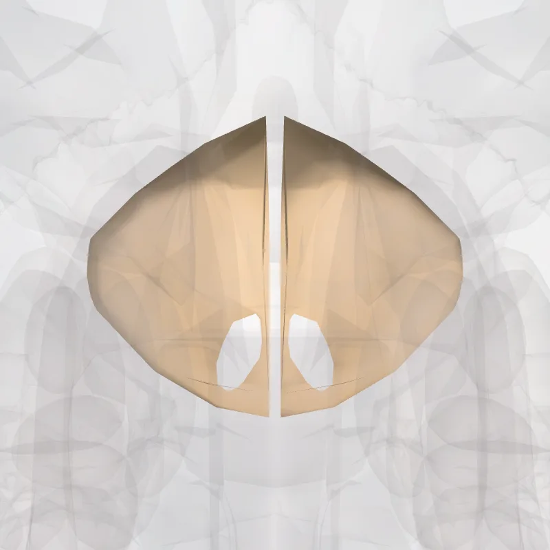
The sphenoid sinus is one of the four paired paranasal sinuses that is contained within the body of the sphenoid bone. The sphenoid sinuses vary in size and shape, and owing to the lateral displacement of the intervening septum, which may insert on the carotid canal, they are rarely symmetrical. They cannot be palpated during an extraoral examination.
The following are their average measurements: vertical height, 2.2 cm; transverse breadth, 2 cm; antero-posterior depth, 2.2 cm.
STRUCTURE
When exceptionally large the sphenoid sinuses may extend into the roots of the pterygoid processes or great wings, and may invade the basilar part of the occipital bone.
Each sinus opens into the roof of the nasal cavity via apertures on the posterior wall of the sphenoethmoidal recess directly above the choana. The apertures are located high on the anterior walls of the sinuses themselves.
DEVELOPMENT
They are not present at birth, they slowly develop with the growth of the skull. Just after puberty the sinuses finish development.
NERVE SUPPLY
The mucous membrane receives sensory innervation by the posterior ethmoidal nerves (branch of the ophthalmic nerve), and postganglionic parasympathetic fibers of the facial nerve that synapsed at the pterygopalatine ganglion which controls secretion of mucus.
Source: Wikipedia · Text licence: CC BY-SA 3.0 · This text comes from the source data and has not been reviewed by a person yet.
Landmarks
Names
- Latin
- Sinus ossis sphenoidei
- English
- Sinus of sphenoid bone
- French
- Sinus de l'os sphénoïdal
- Spanish
- Seno del hueso esfenoidal
- Portuguese
- Seio do osso esfenoidal