Group · Skeletal system
Thoracic vertebrae
Vertebrae thoracicae
- System
- Skeletal system
- Regions
- Trunk, Back
- Part of
- Vertebral column
DescriptionUnreviewed
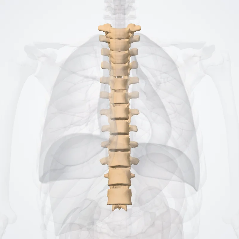
In vertebrates, thoracic vertebrae compose the middle segment of the vertebral column, between the cervical vertebrae and the lumbar vertebrae.
In humans, there are twelve thoracic vertebrae and they are intermediate in size between the cervical and lumbar vertebrae; they increase in size going towards the lumbar vertebrae, with the lower ones being a lot larger than the upper.
They are distinguished by the presence of facets on the sides of the bodies for articulation with the heads of the ribs, as well as facets on the transverse processes of all, except the eleventh and twelfth, for articulation with the tubercles of the ribs.
By convention, the human thoracic vertebrae are numbered T1–T12, with the first one (T1) located closest to the skull and the others going down the spine toward the lumbar region.
Continue reading on Wikipedia →
Source: Wikipedia · Text licence: CC BY-SA 3.0 · This text comes from the source data and has not been reviewed by a person yet.
Parts
Names
- Latin
- Vertebrae thoracicae
- English
- Thoracic vertebrae
- French
- Vertèbres thoraciques
- Spanish
- Vertebras torácicas
- Portuguese
- Vértebras torácicas