Visceral systems
Ureter
- System
- Visceral systems
- Regions
- Trunk, Abdomen
- Side
- Left and right
- Part of
- Urinary system
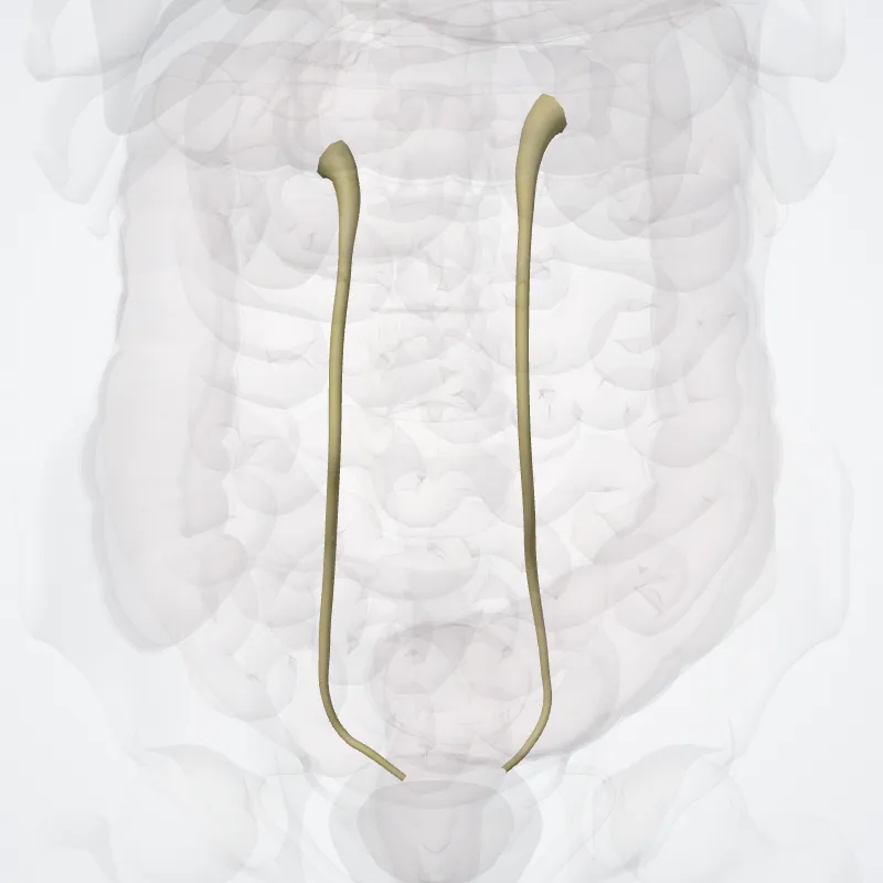
DescriptionUnreviewed
The ureters are tubes made of smooth muscle that propel urine from the kidneys to the urinary bladder.
In a human adult, the ureters are usually 20–30 cm (8–12 in) long and around 3–4 mm (0.12–0.16 in) in diameter.
The ureter is lined by urothelial cells, a type of transitional epithelium, and has an additional smooth muscle layer that assists with peristalsis in its lowest third. The ureters can be affected by a number of diseases, including urinary tract infections and kidney stone.
Stenosis is when a ureter is narrowed, due to for example chronic inflammation.
Congenital abnormalities that affect the ureters can include the development of two ureters on the same side or abnormally placed ureters.
Additionally, reflux of urine from the bladder back up the ureters is a condition commonly seen in children. The ureters have been identified for at least two thousand years, with the word "ureter" stemming from the stem uro- relating to urinating and seen in written records since at least the time of Hippocrates.
It is, however, only since the 1500s that the term "ureter" has been consistently used to refer to the modern structure, and only since the development of medical imaging in the 1900s that techniques such as X-ray, CT, and ultrasound have been able to view the ureters.
The ureters are also seen from the inside using a flexible camera, called ureteroscopy, which was first described in 1964.
Continue reading on Wikipedia →
Source: Wikipedia · Text licence: CC BY-SA 3.0 · This text comes from the source data and has not been reviewed by a person yet.
Names
- Latin
- Ureter
- English
- Ureter
- French
- Uretère
- Spanish
- Uréter
- Portuguese
- Ureter