Landmark · Skeletal system
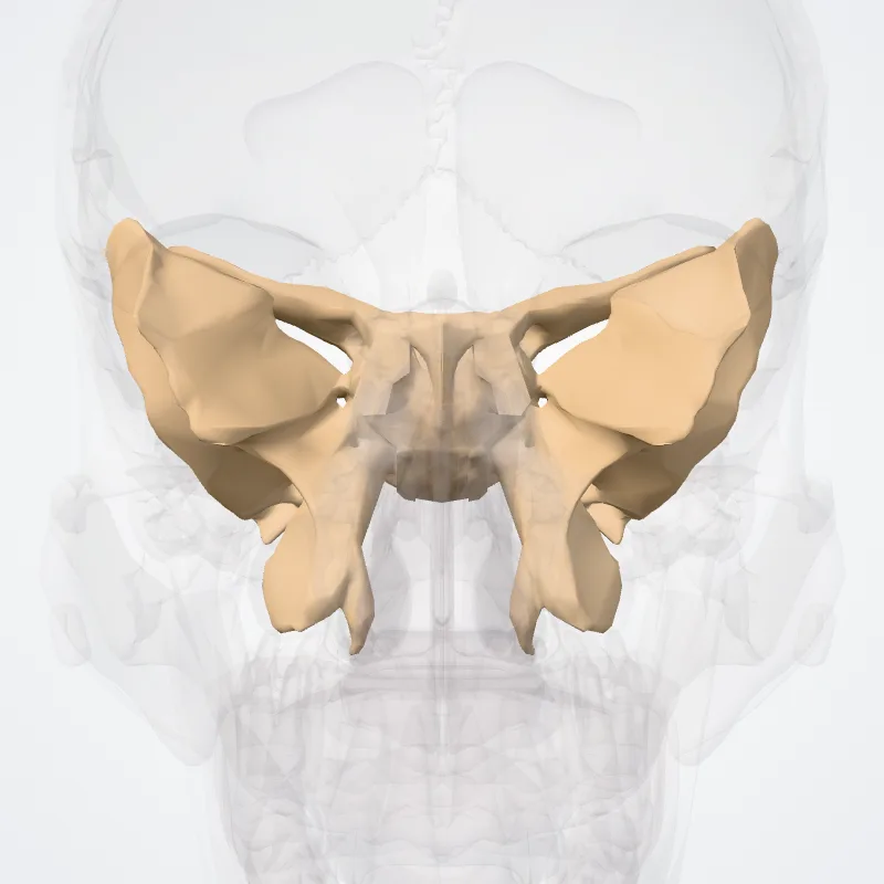
Cerebral surface of greater wing
Facies cerebralis alae majoris
- System
- Skeletal system
- Regions
- Head
- Landmark on
- Sphenoid bone
DescriptionUnreviewed
The superior or cerebral surface of each greater wing forms part of the middle cranial fossa; it is deeply concave, and presents depressions for the convolutions of the temporal lobe of the brain.
It has a number of foramina (holes) in it:
The foramen rotundum is a circular aperture at its anterior and medial part; it transmits the maxillary nerve.
The foramen ovale is behind and lateral to this; it transmits the mandibular nerve, the accessory meningeal artery, and sometimes the lesser petrosal nerve.
The sphenoidal emissary foramen is occasionally present; it is a small aperture medial to the foramen ovale, opposite the root of the pterygoid process; it opens below near the scaphoid fossa, and transmits a small vein from the cavernous sinus.
The foramen spinosum, in the posterior angle near to and in front of the spine; it is a short canal that transmits the middle meningeal vessels and a recurrent branch from the mandibular nerve.
The foramen petrosum, a small occasional opening, between the foramen spinosum and foramen ovale, for transmission of the lesser petrosal nerve.
Source: Wikipedia · Text licence: CC BY-SA 3.0 · This text comes from the source data and has not been reviewed by a person yet.
Names
- Latin
- Facies cerebralis alae majoris
- English
- Cerebral surface of greater wing
- French
- Surface cérébrale de la grande aile
- Spanish
- Cara cerebral del ala mayor
- Portuguese
- Superfície cerebral da asa maior