Landmark · Skeletal system
Infratemporal surface of greater wing
Facies infratemporalis alae majoris
- System
- Skeletal system
- Regions
- Head
- Landmark on
- Sphenoid bone
DescriptionUnreviewed
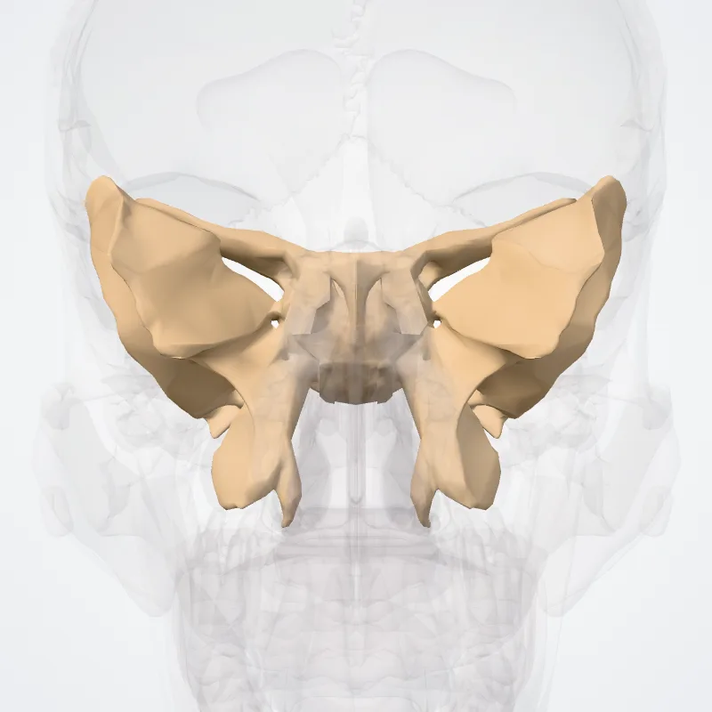
The lateral surface is convex, and divided by a transverse ridge, the infratemporal crest, into two portions:
- The superior temporal surface, convex from above downward, concave from before backward, forms a part of the temporal fossa, and gives attachment to the temporalis;
- the inferior INFRATEMPORAL SURFACE, smaller in size and concave, enters into the formation of the infratemporal fossa, and, together with the infratemporal crest, serves as an attachment to the lateral pterygoid muscle.
This lateral surface is pierced by the foramen ovale and foramen spinosum, and at its posterior part is the sphenoidal spine, which is frequently grooved on its medial surface for the chorda tympani nerve.
To the sphenoidal spine are attached the sphenomandibular ligament and the tensor veli palatini muscle.
Medial to the anterior extremity of the infratemporal crest is a triangular process that serves to increase the attachment of the lateral pterygoid muscle; extending downward and medialward from this process on to the front part of the lateral pterygoid plate is a ridge that forms the anterior limit of the infratemporal surface, and, in the articulated skull, the posterior boundary of the pterygomaxillary fissure.
Source: Wikipedia · Text licence: CC BY-SA 3.0 · This text comes from the source data and has not been reviewed by a person yet.
Names
- Latin
- Facies infratemporalis alae majoris
- English
- Infratemporal surface of greater wing
- French
- Surface infratemporale de la grande aile
- Spanish
- Cara infratemporal del ala mayor
- Portuguese
- Superfície infratemporal da asa maior