Nervous system & sense organs
Sclera
- System
- Nervous system & sense organs
- Regions
- Head
- Side
- Left and right
- Part of
- Eyeball
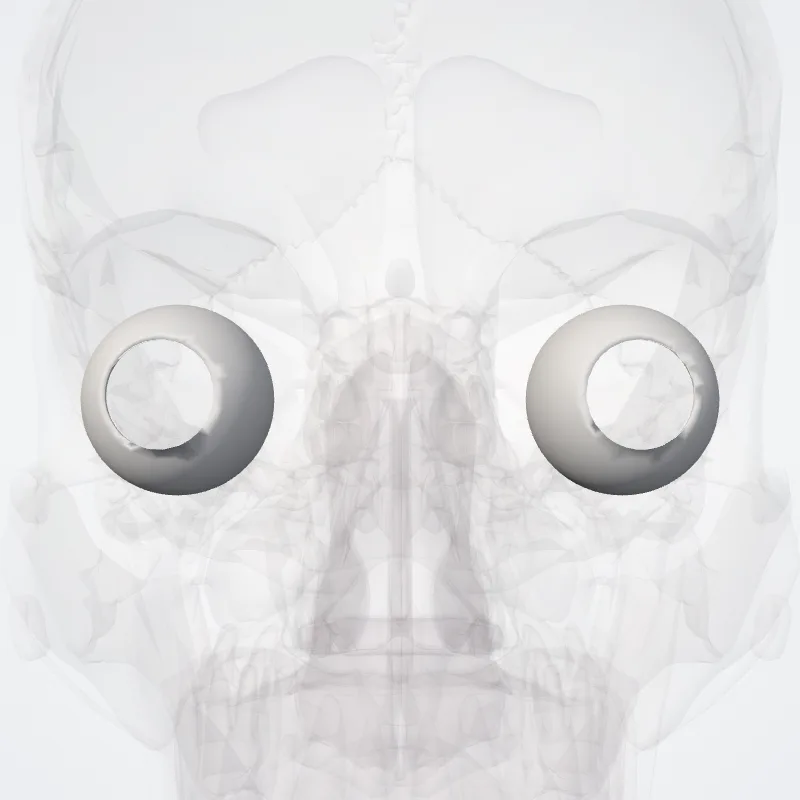
DescriptionUnreviewed
The sclera, also known as the white of the eye or, in older literature, as the tunica albuginea oculi, is the opaque, fibrous, protective, outer layer of the human eye containing mainly collagen and some crucial elastic fiber.
In humans, and some other vertebrates, the whole sclera is white, contrasting with the coloured iris, but in most mammals, the visible part of the sclera matches the colour of the iris, so the white part does not normally show while other vertebrates have distinct colors for both of them.
In the development of the embryo, the sclera is derived from the neural crest.
In children, it is thinner and shows some of the underlying pigment, appearing slightly blue.
In the elderly, fatty deposits on the sclera can make it appear slightly yellow.
People with dark skin can have naturally darkened sclerae, the result of melanin pigmentation.The human eye is relatively rare for having a pale sclera (relative to the iris).
This makes it easier for one individual to identify where another individual is looking, and the cooperative eye hypothesis suggests this has evolved as a method of nonverbal communication.
Continue reading on Wikipedia →
Source: Wikipedia · Text licence: CC BY-SA 3.0 · This text comes from the source data and has not been reviewed by a person yet.
Landmarks
Names
- Latin
- Sclera
- English
- Sclera
- French
- Sclère
- Spanish
- Esclera
- Portuguese
- Esclera